
The Wrist:
The wrist is a complex joint that connects the radius and ulna (the 2 bones in the forearm) to the carpals in the hand. In most textbooks you'll find the wrist's purpose stated as: "The wrist (and hand) allow for the manipulation of objects in space and provide us with the dexterity required for fine motor skills." While that is the case for most people, acrobats & gymnasts often use this joint as a weight bearing structure. Sadly, it's not nearly as "beefy" as the foot and ankle, so injury risk is higher and maintenance exercises are essential.
Osteology of the Wrist:
The wrist is the junction of the distal end of the radius/ulna and the adjacent carpal bones. It is often compared to the ankle joint(s) in structure, however through evolution, the wrist has become more delicate and lost many of the characteristics that would allow it to be a truly effective weight-bearing joint. The bones are smaller, there is less cartilage, and the ligaments are thinner - a trifecta of risk for injury and instability.

Palmar view of the carpal bones
The eight carpal bones can be divided into two rows of four bones. The proximal (or closer) row is composed of: (running from thumb to pinky) the scaphoid bone, the lunate bone, the triquetrial bone, and the pisiform bone. The distal (or farther) row is composed of: the trapezium bone, the trapezoid bone, the capitate bone, and the hamate bone. Why are the names important? They're not (unless you're heading into the medical field) - but it helps to know so that I can tell you that the scaphoid is the most commonly fractured of the bones as its location makes it more susceptible to weight and force.
Though there is no direct connection between the carpals and the end of the ulna, there is a cartilaginous disc (shown in the picture below) that acts to allow increased congruence between the ulna and the carpal, as well as maintain the space on the ulnar side of the joint. This disc is known as the Triangular Fibrocartilage Complex (or TFCC) and is commonly injured in weight bearing activities. Basically, the TFCC serves as the "meniscus of the wrist" and like the meniscus, can be compressed, torn and damaged when there is weight on the wrist and a twisting motion (think handstands that fall or a pivot on the wrist around the trapeze). The TFCC has connective tissue attachments onto the radius as well (through ligaments/fascia) so it's worth noting that it can be involved in many types of wrist problems.

One reason that the wrist is so complicated is that every small bone forms a joint with the other small bones adjacent to it (aka "Intercarpal Joints"). This means that technically, there are "dozens" of small joints in the wrist that assist in allowing the versatility of movement that our hands and wrists require to function. Articular cartilage covers the ends of each of the bones where they meet in a joint, thus improving glide and protecting joint integrity. It wouldn't be reasonable to breakdown all of the joints - rather just recognizing that they exist is sufficient for the goals of this post.
For our purposes, we need only look at three of the more functional joints in the wrist:
- The distal radioulnar joint: This is the point just prior to the wrist where the radius and ulna connect. This is not a synovial joint, but rather exists because of the interosseus membrane (a thick ligamentous structure that connects them). This is best described as a "pivot" joint and allows (in conjunction with the proximal radioulnar joint at the elbow) the forearm to pronate (turn down) and supinate (turn up). The stability of this joint is essential for any weight bearing on the hand and wrist as it creates the "mortice" for the proximal carpals.
- The radiocarpal joint: This is the intersection of the distal end of the radius and the proximal row of carpals (as described above). It is a true synovial joint, meaning that there is a joint capsule containing lubricating fluid within. The proximal row of carpals is convex on the concave radius. This joint allows the hand and wrist to move down (palmar flexion) and up (extension or dorsiflexion). In gymnastics/acrobatics/yoga, more extension is required than most other sports as weight bearing forces the wrist into that position. For this reason, often the radiocarpal joint is hypermobile (extra flexible), bordering on unstable, and is very susceptible to injury. This joint also allows a side to side glide which causes the hand and wrist to "tilt" left and right into radial deviation and ulnar deviation.
- The midcarpal joint: This is the intersection of the proximal row and the distal row of carpals. While not an Isolated single joint, the midcarpal joint is essential for achieving end-range motion into extension (something acrobats require). There is not as much motion available in this "joint" as the radiocarpal, but these bones glide to allow to the same motions as the radiocarpal joint. Think of the joint as a "helper joint" in that alone it cannot perform the motions, but rather boosts the radiocarpal joint's range.
The Ligaments of the Wrist:
In the wrist, the ligaments are the passive supports that ensure that the structure of the joint is maintained. There are many ligaments in the wrist joint, and rather than name them all, we can group them into two categories:
Dorsal Intercarpal Ligaments: A series of ligaments that extend transversely across the dorsal (back) surface of the wrist connecting the carpals to one another and the carpals to the radius/ulna.
Dorsal Ligaments
Palmar Intercarpal Ligaments: A series of ligaments that extend transversely across the palmar (front or volar) surface of the wrist connecting the carpals to one another and the carpals to the radius/ulna. These ligaments are thicker than the dorsal ligaments (which works our well for us as these ligaments support the front of the wrist and palm in weight bearing). Two named ligaments exist: the palmar (volar) radiocarpal ligament and the palmar (volar) intercarpal ligament.
Palmar Ligaments (termed Volar Ligaments)
There are additional ligaments that assist in attaching specific carpals to one another, but for our purposes are not necessary to discuss. (If you have specific questions about the ligaments, please email me or post a question in the comments section).
Muscles of the Wrist:
Similar to the muscles that control the movement of ankle, the wrist and hand muscles are located primarily in the forearm. Often they work in groups (aka synergistically) to allow for the dexterity and movement of these joints.
The tendons of these muscles are longer from the end of the muscle belly to the insertion, and thus require two thick bands at the wrist to anchor them in place:
The anterior band, known as the Flexor Retinaculum (aka transverse carpal ligament), travels across the palmar side of the wrist and forms the "Carpal Tunnel." This tunnel is supported on the sides and back by the carpal bones, and has all of the flexor tendons (in synovial sheaths), the median nerve, and several arteries and veins traveling through it. It is negatively affected by inflammation of prolonged pressure and is a commonly injured area (think carpal tunnel syndrome).

The posterior band, known as the Extensor Retinaculum anchors the extensor tendons and assists in supporting the posterior structure of the wrist.
Alright, on to the muscles...In order to simplify a rather massive amount of information, I've divided these muscles into the action they perform.
Anterior and Posterior views of the muscles that control the wrist and hand
Flexion: bending the wrist towards the palm. Muscles include: Flexor Carpi Ulnaris (prime mover), Flexor Carpi Radialis (prime mover), Palmaris longus (tightens the skin and fascia in the palm), Flexor Digitorum Superficialis (finger flexor, assists only).
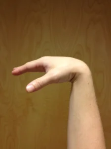
Wrist flexion (right hand)
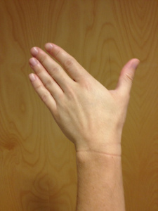
Extension: bending the wrist backwards; the primary position for weight bearing. Muscles include: Extensor Carpi Radialis Longus (prime mover), Extensor Carpis Radialis Brevis (prime mover), Extensor Carpi Ulnaris, Extensor Digitorum (assists only).

Wrist extension (right hand)
Radial Deviation: tilting the hand and wrist toward the thumb and radius. Muscles include: Flexor Carpi Radialis & Extensor Carpi Radialis (they work together to cancel the flexion and extension and instead just pull toward the radius). This motion is more important in weight bearing activities like those performed on the balance beam, rings, pommels, and p-bars when using the wrists to maintain the body an inverted position.

Radial Deviation (left hand)
Ulnar Deviation: tilting the hand and wrist toward the pinky and ulna. Muscles include: Flexor Carpi Ulnaris & Extensor Carpi Ulnaris (they work together to cancel the flexion and extension and instead just pull toward the ulna). This motion is more important in weight bearing activities like those on the balance beam, rings, pommels, and p-bars when using the wrists to maintain the body in an inverted position.
Ulnar Deviation (left hand)
The anatomical complexity and versatility of the wrist not only allows us all the dexterity to function, it also allows acrboats, gymnasts and yogis many of the abilities required for skill acquisition in their sport.
References:
- Dutton, M. (2008). Orthopaedic: Examination, evaluation, and intervention (2nd ed.). New York: The McGraw-Hill Companies, Inc.
- Levangie, P.K. and Norkin, C.C. (2005). Joint structure and function: A comprehensive analysis (4th ed.). Philadelphia: The F.A. Davis Company.
- Neumann DA. Kinesiology of the musculoskeletal system: Foundations for Physical Rehabilitation.2nd Ed. Elsevier Health Sciences; 2009
- Parmelee-Peters, K., Eathorne, S. (2005). The Wrist: Common Injuries and Management. Primary Care, Clinics in Office Practice, 35-70.
- Wadsworth, C., The wrist and hand examination and Interpretation, J. Orthopedic and sports physical therapy, 1983, 108-20.